作品賞
纖毛蟲與高鐵來比快
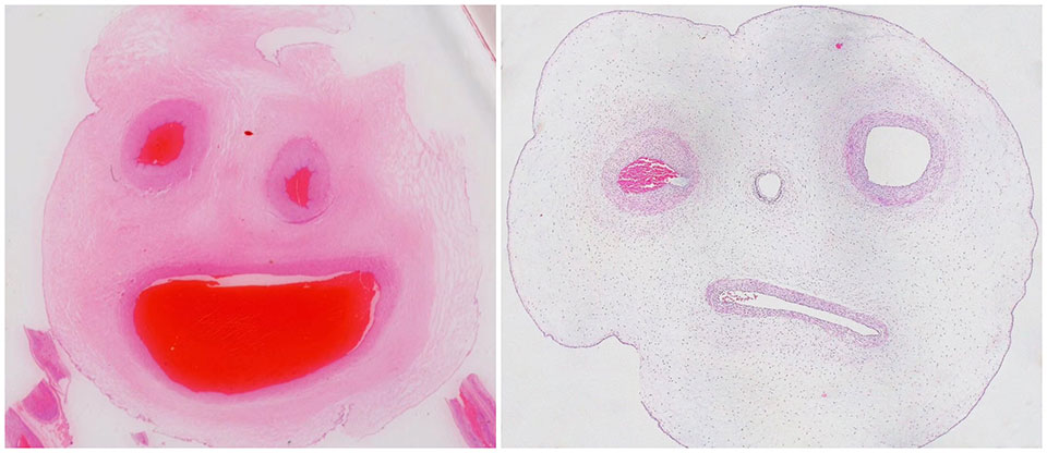
圖片為臍帶組織切片HE染色顯微照片。臍帶是胎兒和母親間特殊而唯一的紐帶。形狀如繩索,表面光滑透明,內含結締組織和一支臍靜脈,一對臍動脈。臍靜脈沿著胎兒腹壁內面通過肝的血竇、臍動脈與胎兒主動脈相通連。圖中臍帶組織切片通過HE染色在顯微鏡中呈現出嬰兒般的兩張臉。臉上的兩隻“眼睛”正是臍動脈,而“嘴巴”正是臍靜脈。
The photo is an HE staining micrograph of umbilical cord tissue section. The umbilical cord is a unique and special bond between the fetus and the mother. Shaped like a rope, its surface is smooth and transparent, containing connective tissue and a umbilical vein, and a pair of umbilical arteries. The umbilical vein is connected to the fetal aorta through the sinuses of the liver and the umbilical artery along the inner surface of the abdominal wall of the fetus. In the picture, the umbilical cord tissue section displays two baby-like faces under the microscope as the result of HE staining. The two "eyes" on the face are the umbilical artery, and the "mouth" is the umbilical vein.
2017年5月22日拍攝於上海市 衡道病理-上海交通大學個性化醫學研究院 智慧病理聯合實驗室。拍攝使用的顯微鏡為Leica Microsystems CMS GmbH Ernst-Leitz-Str. Model DMIL LED Fluo.臍帶組織切片經過HE染色在顯微鏡下拍攝。
Taken on May 22, 2017 in Shanghai, HengDao Histo Pathology Diagnostic Center – Institute of Personalized Medicine of Shanghai Jiao Tong University (SJTU) Joint Laboratory of Intelligent Pathology. The microscope used was Leica Microsystems CMS GmbH Ernst-Leitz-Str. Model DMIL LED Fluo. Umbilical cord tissue sections were photographed under the microscope after HE staining.